Understanding Vision.
How The Eye Works
The human eye is truly amazing. It focuses light onto
the back of your eye to form images or "pictures", much like a camera exposing film. The eye instantly changes these images
into electrical signals and sends them to your brain. The brain interprets
the signals and you experience "seeing".
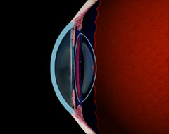
The basic structures
of the eye responsible for vision are:
-
Cornea: This clear outer lens provides two-thirds of the
focusing power of the eye. The cornea is made
up of transparent tissue, which allows light to pass through. The cornea
focuses the light by bending it so the light rays form
an image on the retina. Since the cornea has the greatest bending (focusing)
power, it is the cornea's shape that determines a great deal of quality of
your vision.
-
Iris & Pupil: The colored part of the eye is called the iris
and functions much like the iris of a camera, opening and closing, to control
the amount of light entering through the pupil (that dark opening
in the center the iris).
-
Crystalline lens: The crystalline lens is located behind the iris
and provides one third
of the focusing power of the eye. The crystalline lens works to further
bend light rays as they pass through the eye to form an image on the retina.
-
Retina: Located in the lining at the back of the eye, the retina
acts as an electrical system to send impulses to your brain via the optic
nerve. The retina
contains photoreceptor cells that collect information from light as it
passes through the cornea and crystalline lens to the back of the eye.
Your brain interprets the retina's electrical response into what you to experience
as images.
-
Fovea: The focal point at the center of the retina is called the
fovea. Light focused here produces the sharpest
vision.
The
eye focuses by bending incoming light rays to meet at a single
point. Ideally this single point lands directly on the fovea, the central point
of the retina. If the light rays reach this perfect placement, you experience
clear, sharp images. However, if the focal point is behind the retina or in
front of the retina, the image on the retina will not be fully formed and will
be interpreted by your brain as blurred. This is very much like focusing a
projector onto a blank movie screen. If the projection is too close or too
far from the screen, the images are blurred. Set at the correct distance, you
may enjoy the show!
When you have an eye examination at Kindermann Eye, your eye doctor is able
to determine whether you are nearsighted, farsighted, and/or astigmatic. During
your eye exam, it will be determined where your eye focuses light.
Depending on your refraction, your eye doctor will discuss
different treatment options with you.
| |
Normal vision:
The cornea is able to bend light rays to focus directly onto your retina.
Both
near and far vision is satisfactory.
|
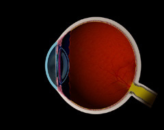
Light focuses at a single point on the retina.
(Move mouse over image.)
|
| |
Nearsightedness (myopia):
The eye is too long (note the shape of the
cornea) and the focal point is in front of the retina.
Distant objects become
blurred, while near vision is usually satisfactory.
|
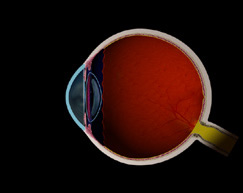
Light focuses at a point in front of the retina.
(Move mouse over image.)
|
| |
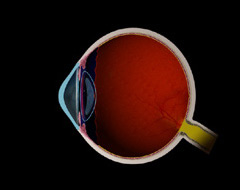
Farsightedness (hyperopia):
The eye is short (note the shape of
the cornea) and the focal point is behind of the retina.
Distance vision
is typically satisfactory, with difficulty occurring at reading distance.
|
Light focuses at a point behind the retina.
(Move mouse over image.)
|
| |
Astigmatism:
The astigmatic cornea is shaped
irregularly, like a football, having an oval rather than round curvature.
Light rays do not come to a single focus point, but rather objects are focused
at more than one point, distorting both distance and near vision.
|
Light focuses on more than one point.
(Move mouse over image.)
|
| |
Presbyopia:
A natural part of the aging process, presbyopia occurs
when a person is unable to focus on near objects because of insufficient accommodation
ability.
Accommodation is the
ability of your natural (crystalline) lens to change from distance vision to
near vision as desired.
Presbyopia begins during the early to mid-forties, and manifests as an inability
to read without glasses.
|
Accommodation is the ability of the lens to
move between close
and distance vision.
(Move mouse over image.)
|
|
|
 (856) 667-3937 Kindermann Eye Associates
3001 Chapel Avenue, Suite 200
Cherry Hill, New Jersey 08002 |
Eye Doctor South Jersey Kindermann Eye Associates - 3001 Chapel Avenue, Suite 200, Cherry Hill, New Jersey 08002 Excellence in eye care for patients seeking a quality caring eye doctor in South Jersey. Dr. W Reed Kinderman is a premier ophthalmologist, New Jersey eye surgeon, specializing in cataract surgery, refractive surgery, laser guided cataract surgery, Tecnis multifocal lens implant, ReStor intraocular lens, Crystalens, Toric intraocular lens, glaucoma, strabismic eye muscle disorders, ophthalmology, and the full spectrum of eye care in New Jersey, Delaware Valley, Philadelphia, Mullica Hill, Ashland, Echelon, Thorofare, Riverton and surrounding areas. |
Copyright Kindermann Eye Associates, All Rights Reserved.
|

